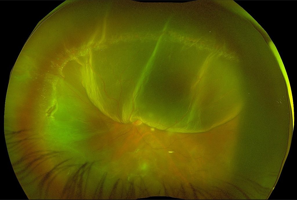
33 year old male patient came with sudden diminision of vision in left eye since 3 days . He was a high myope and had history of previous LASIK and cataract surgery in both eyes . On examination his Best corrected visual acuity in left eye was CF-CF (counting fingers close to face ). Anterior segment showed pseudophakia . Fundus examination showed Subtotal Rhegamtogenous retinal detachment with horse shoe tear and multiple lattices with holes . Patient was advised and underwent 27g parsplana vitrectomy with silicon oil injection.
Preoperative fundus photo of left eye
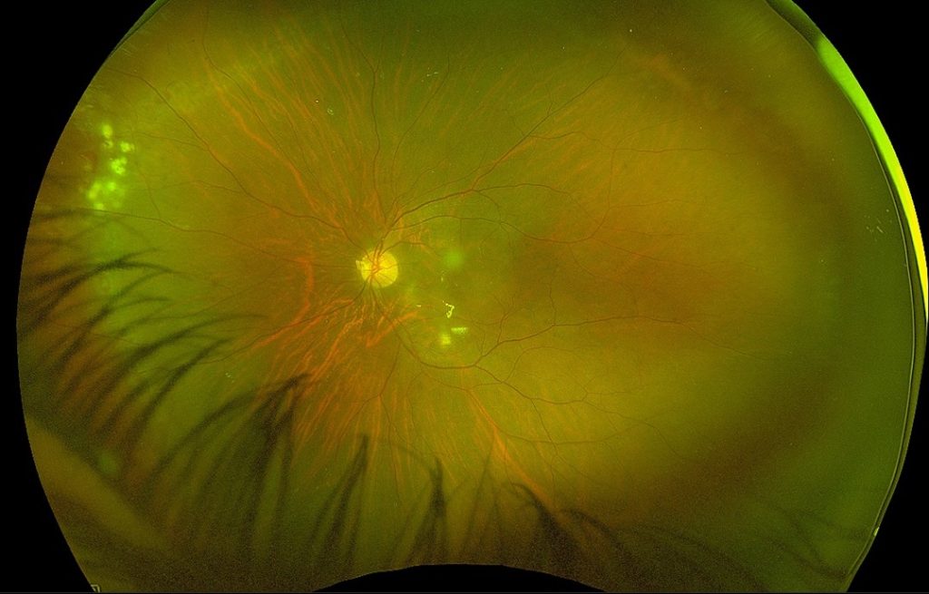
Postoperative fundus photo with silicon oil filled globe and attached retina
Total Eye Care For All
We offers a complete range of eye care services to All.